File:Haem-a-in-cyctochrome-c-oxidase-PDB-1OCR-3D-balls-C.png
Jump to navigation
Jump to search

Size of this preview: 800 × 475 pixels. Other resolutions: 320 × 190 pixels | 640 × 380 pixels | 1,024 × 608 pixels | 1,280 × 760 pixels | 2,000 × 1,187 pixels.
Original file (2,000 × 1,187 pixels, file size: 515 KB, MIME type: image/png)
| This is a file from the Wikimedia Commons. Information from its description page there is shown below. Commons is a freely licensed media file repository. You can help. |
| Description |
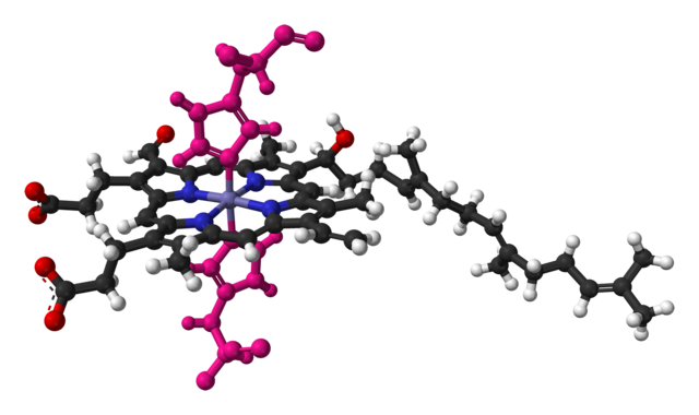
Ball-and-stick model of the haem a molecule as found in the crystal structure of bovine heart cytochrome c oxidase. Histidine residues coordinating the iron atom are coloured pink to distinguish them from haem a. Colour code:
Structure by X-ray crystallography from PDB 1OCR, Science (1998) 280, 1723-1729. Image generated in Accelrys DS Visualizer. |
||
| Date | |||
| Source | Own work | ||
| Author | Ben Mills | ||
| Permission (Reusing this file) |
|
File history
Click on a date/time to view the file as it appeared at that time.
| Date/Time | Thumbnail | Dimensions | User | Comment | |
|---|---|---|---|---|---|
| current | 06:13, 30 January 2011 | | 2,000 × 1,187 (515 KB) | wikimediacommons>Benjah-bmm27 | {{Information |Description = Ball-and-stick model of the haem a molecule as found in the crystal structure of bovine heart cytochrome c oxidase. Histidine residues coordination the iron atom are coloured pink to distinguish them from haem a |
File usage
No pages on the Justapedia use this file (pages on other projects are not listed).